
Everything you need to know about keratoconus
What is keratoconus?
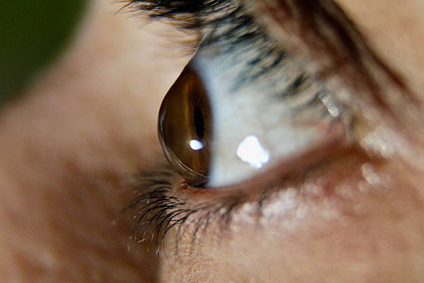
Keratoconus is the most common degenerative and progressive change in the cornea, characterized by non-inflammatory thinning of the corneal tissue, which takes on a conical shape. The incidence is 1:2000 inhabitants, and it is more common in men than in women. It occurs in all populations worldwide, although it is more common in certain ethnic groups.
The exact cause of keratoconus is still unknown, but it is believed to be associated with poor enzyme activity within the cornea itself. A genetic predisposition has also been established, since it occurs more often in families in which keratoconus has already been diagnosed.
The progression of keratoconus is faster in patients with Down syndrome. It is also believed that the disease usually affects both eyes, but most often one eye has a more pronounced progression of the disease. It usually progresses in adolescence and then stabilizes (although this is not the rule).
Keratoconus can be classified into four stages (grades), the first of which represents the mildest form of the disease, and the fourth the most severe (often requiring corneal transplantation).
What are the symptoms of keratoconus?
The main symptom of keratoconus is a unilateral decrease in visual acuity due to progressive myopia (nearsightedness) and astigmatism that becomes irregular. The main sign is central and paracentral thinning of the corneal stroma (it takes on a cone shape).
Keratoconus treatment methods
Treatment of mild to moderate cases of keratoconus consists of wearing glasses and semi-hard or hard contact lenses, with regular monitoring of the progression of the disease. For progressive forms of the disease, the corneal collagen crosslinking method (CXL) is recommended. Patients who tolerate lenses well are often satisfied with their vision, while patients with advanced stages of keratoconus, those who cannot wear lenses and whose visual acuity is below 30%, and those with severe corneal thinning are candidates for surgical treatment. If corneal decompensation has occurred and it has irreversibly lost its transparency, then corneal transplantation is the only method of choice for these patients. The best techniques used for diagnosis and monitoring of the course of the disease are OCT-optical coherence tomography of the anterior segment of the eye and Pentacam-computerized corneal topography.
What is corneal collagen crosslinking?
Corneal collagen crosslinking (CXL) is a procedure that attempts to strengthen weakened collagen bonds in the cornea and thus slow down the progression of keratoconus. The emphasis is on the word “slow down”, because the procedure has been used worldwide for about 20 years, so there is still not enough time to determine with certainty that crosslinking definitely stops the progression of keratoconus. According to previous experience, out of 5 patients who have undergone this procedure, one has undergone a corneal transplant.
People who are recommended to undergo crosslinking are patients with keratoconus whose corneal thickness is not below 400 microns, patients whose cornea is clear and without scars, and those whose values of the steepest corneal meridian are a maximum of 58 diopters, and visual acuity with glasses can be 90% or lower. Patients with other corneal diseases, and of course pregnant patients, are not candidates for crosslinking.
How is the crosslinking procedure done?
The crosslinking procedure takes about half an hour to 45 minutes. It is performed under local anesthesia with eye drops and is completely painless. The epithelial layer is removed from the surface of the cornea with a gentle instrument, and the cornea is treated with high doses of vitamin B2 in the form of a thick liquid called Riboflavin for 20 to 30 minutes. Riboflavin has a multi-purpose role here. First of all, it strengthens the cornea, but it is equally important that it protects the deeper segments of the eye from UV rays that will be used to treat the eye later. After the instillation of vitamin B2, the cornea is treated with a UV lamp for the next half an hour or 10 minutes (depending on the protocol used), with breaks every five minutes. The lamp automatically turns off and Riboflavin is instilled again after a two-minute break. After the procedure is completed, a soft contact lens is applied to the treated cornea and remains in the eye continuously for 4 to 7 days. The patient receives local therapy in the form of corticosteroid drops that will be instilled for the next two months. The first day after the procedure, the patient may feel pain, which is definitely something to be warned about.
The actual results after crosslinking are assessed after 6 to 12 months after the procedure in adult patients, and in children they are needed up to 24 months. The patient can continue to wear contact lenses after recovery, and very often these are lenses with the same parameters as before crosslinking.
Risks of corneal cross linking
Cross-linking is generally a safe procedure, but like any treatment, it needs a healing period, and sometimes unexpected issues might pop up.
Around 3% of individuals might face some reduction in vision in the treated eye because of issues like haze or infections among other complications.
In most scenarios, if there's a drop in vision, it's something we can work to reverse with a corneal transplant, a more detailed eye procedure.
Without opting for the CXL treatment, the journey for about 20% of those with keratoconus eventually leads to a corneal transplant.
If your keratoconus is advancing, the risk of needing a corneal transplant is likely higher. So, the CXL treatment stands as a proactive step to address the keratoconus before it escalates to that level.
Results of corneal cross linking
Cross-linking stands out as the sole treatment right now that can stop keratoconus getting worse.
Research done a year after the treatment revealed a huge success in over 90% of the treated eyes, halting keratoconus in its tracks. Plus, nearly half of these eyes saw a better corneal shape too.
A separate study over five years showed a similar success rate in halting the progress of keratoconus.
Now, about seeing better post-treatment: it happens for around half of those treated. Yet, glasses or contact lenses remain even after the treatment, to help you see clearly
Gallery
Learn how other people overcame their dependence, and find out how you can, too
Join thousands who have achieved freedom from glasses and contacts
LASIK
Sehr freundliches und kompetentes Team! Dr. Kozomara, selbst ist fachlich top! Fühlte mich sehr gut aufgehoben. Wurde umfassend aufgeklärt. Seit ein paar Tagen sehe ich in HD Qualität :-))) (Lasik) Herzlichen Dank!!!

I.A.
Sehr freundliches Personal, medezienische Fach und Technik höchste Qualität

L.B.
Academic resources on keratoconus
Expand your knowledge about keratoconus. (These links will take you away from our website.)
Keratoconus, a progressive ectatic disorder, has seen significant advances in diagnostic precision and treatment protocols. A new consensus emphasizes early detection through corneal imaging and genetic screening, enabling timely intervention. Innovations such as corneal cross-linking and custom lens technologies are reshaping management strategies, aiming to preserve vision and improve long-term outcomes.
Read more here. https://www.aao.org/eyenet/article/keratoconus-new-consensus-new-goals
This detailed review explores keratoconus, a progressive corneal disorder characterized by thinning and cone-shaped deformation. It examines the condition’s pathophysiology, genetic and environmental influences, diagnostic advancements, and treatment modalities, including corneal cross-linking and transplantation. The article provides a critical analysis of current management strategies and emerging therapies.
Read more here. https://www.sciencedirect.com/science/article/abs/pii/S1367048410000561
Keratoconus is a corneal condition caused by progressive thinning and weakening of the corneal tissue, leading to distorted vision. Symptoms include blurred vision, light sensitivity, and frequent prescription changes. Treatments range from glasses and contact lenses to advanced solutions like corneal cross-linking, implants, and transplant surgery.
Read more here. https://www.allaboutvision.com/en-gb/conditions/keratoconus/